Endodontics (Root Canal Treatment)

A tooth can become painful when the nerve (pulp) becomes inrreversibly inflammed due to the following reasons :
[list style=”style4″ line=”no”]
- Deep dental caries
- Trauma
- Crack or chip in the tooth
- Repeated dental procedures on the tooth
[/list]
Signs and symptoms that tooth require a root canal treatment are as follow:
[list style=”style4″ line=”no”]
- Toothache
- Prolonged sensitivity to heat or cold
- Painful to touch or chewing
- Discoloured tooth
- Swelling
[/list]
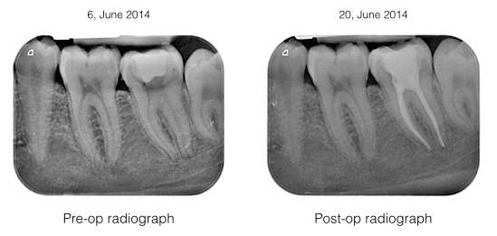
Examination: Lower left second molar has a gross decay. The tooth was very tender to percussion. There was no swelling noted. Tooth was painful to airblow. Intra-oral periapical radiography showed small periapical lesion on both mesial and distal root with widening of the lamina dura.
Diagnosis: Acute apical periodontitis on tooth 37
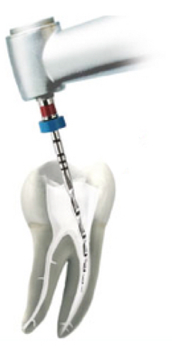
Treatment: Tooth 37 was anesthetized and filled. Root canal treatment (RCT) on tooth 37 was initiated under rubber dam isolation. Microscopic investigation did not reveal any fracture. All 3 canals were shaped, cleaned, dried and dressed with calcium hydroxide for a week. Tooth 37 was subsequently obturated up to full working length.

Examination: Tooth 36 has a large restoration. The tooth is mildly tender to percussion and not resposive to cold test. Radiograph showed the tooth with a small periapical lesion. Since the tooth does not cause much trouble at the time of examination, patient requested, to observe the condition further.
6 months later, the pain on the lower left 1st molar returns and the radiograpgh showed the periapical lesion has increased in size. Tooth was very tender to touch.
Diagnosis: Acute apical periodontitis of tooth 36
Treatment: Root canal treatment was carried out on tooth 36 under rubber dam isolation. Both mesial canals are curve. All 3 canals were cleaned and shaped. Calcium hydroxide dressing were placed for disinfection. One week review showed tooth 36 is no longer painful. Obturation was carried out on all the 3 canals.

Examination : Tooth 47 has a large overhanged composite restoration, and slight swelling on gum. The tooth was very tender to percussion. Cold test and pulp test were negative. Radigraph showed tooth 47 has overhanged and leaking filling. Someform of root theraphy was initiated but the root canals have not been shaped, cleaned and filled.
Diagnosis : Acute apical periodontitis 47 with previously attempted root therapy.
Treatment : Old composite filling was replaced to to prevent any further ingression of microorganism into the root canal system. RCT was initiated under rubber dam isolation. Under microscope, there was one mesio lingual canal and a C-shape canal extending from mesiobuccal to distal. All canals were shaped with rotary nickel-titanium files and cleaned with ultrasonic irrigation. Canals were dressed with calcium hydroxide. The tooth was asymptomatic after 3 weeks review and was subsequently obturated to full working length. A full gold crown restoration was placed to prevent any coronal leakage.

Examination : Tooth 17 has a large old amalgam restoration. Bite test was positive whereby pain was noted upon releasing of bitting pressure. Cold test is positive suggesting the tooth is still vital. There was a hairline crack noted under microscope upon removing of old amalgam filling
Diagnosis : Acute apical periodontirs 17 due to crack tooth.
Treatment : Old amalgam restoration was replaced with composite filling and RCT was carried out under rubber dam isolation. Under microscope, all 4 root canals (MB1, MB2, DB, P canals) were located shaped, cleaned and obturated up to full working length. The tooth was subsequently restored with a semi precious metal crown.


